

Capture Every Detail with Moticam Pro S7 MONO Featuring Sony Pregius Sensor
Motic America
Read Article
Free Ground Shipping on Orders Over $300 Across USA and CANADA
Posted by Motic America on

Introduction
Beer is one of the oldest beverages (M. Nachel, and S. Ettlinger, 2012; T. Coultate ,2016). The earliest evidence of beer produced from barley dates to about 3500–3100 BC, from the site of Godin Tepe in the Zagros Mountains of western Iran (Wikipedia). Today microscopes play an important role in the brewing of beer and wine. The primary use of a microscope is for counting yeast cells using a special counting chamber called a hemocytometer to determine viability of the yeast. Yeast cell counts are used to determine the pitching rate and yeast cell density when bottling beer and determining yeast density in sediment. During mashing, the malt is mixed with water and enzymes (amylases) to convert insoluble starch into soluble sugars. The liquid is called wort which is boiled and yeast is added so fermentation can start. Pitching is a term brewer’s use for adding yeast to wort “beer starter”. During fermentation yeast converts sugars from the malt into carbon dioxide and ethanol. Yeast collects at the bottom of the vessel and is later removed. Young beer is cooled and stored for a few months allowing the flavor to mature. Once yeast has been pitched and removed it is considered beer.
Yeast is a single celled fungi used in baking, wine, beer fermentation and basic cell biology research. Yeast cells are small about 0.010 mm (10 microns) and require a microscope to see and count them. A microscope for amateur or professional brewers allows them to count the yeast, detect infection by bacteria and other filamentous fungi (mold).
Microscopes for Viewing Yeast used in making Alcoholic Beverages
To see and count yeast a basic light microscope is required (Microscopy for the Winery - UC Davis). The microscope should have at least 10, 20 and 40X objectives and 10X eyepieces which will let you see the yeast cells and bacteria. Better microscopes will also have 4X and 100X (oil immersion) objectives (Microscope use in Brewing). Whichever microscope is used it should have coarse and fine focus knobs, a bright light source, an Abe or Phase contrast condenser. A microscope trinocular head is good if documenting observations with a camera is required. A mechanical stage with x, y controls to move glass slides or the counting chamber containing a grid for counting yeast is useful. For those that want to study the internal structure of yeast cells a 100X objective is valuable.
For professional use the Motic BA310E Phase contrast microscope or BA410E would be ideal.
For amateurs a Motic BA210E would be a good solution


Determining Viability with Methylene blue and a Hemocytometer Slide
To count yeast cells a hemocytometer slide with a grid etched on the glass is used. These slides cost between $35 and $150 and are readily available online. In addition you will need some small pipettes and a dye to determine yeast cell viability. Typically methylene blue or Trypan blue dye is used. Mix 0.1% w/v methylene blue with your yeast solution 1:1. Some users prefer Trypan blue (https://en.wikipedia.org/wiki/Trypan_blue). To learn more about hemocytometers and how to use them see YouTube video links below and/or Wikipedia. Cells that stain blue are dead, while those that are unstained are alive. You should standardize the time when you count the cells in stain as more cells will stain blue the longer the cells are in stain.

Hemocytometer slides have two sections with grids etched into the glass. The grids are divided into smaller squares of known volume. The cells are counted in the squares, those cells found on the top and left grid lines of the outer square are ignored. Usually cells in the top and bottom compartments are counted and averaged. If the difference in the count is more than 2X you should repeat the test and make sure the yeast cells are well dispersed. You may have to dilute the samples before counting.
These chambers are also used for counting blood cells (hence term Hemocytometer), sperm, plankton, bacteria or any other type of small particulate matter in suspension. A simple Tally counter is helpful for counting the yeast.
To detect infection in beer often agar dishes with selective growth medium are used as the number of bacteria is usually low.

Grid on a slide with a counting chamber. Hemocytometer grid: red square = 1.0000 mm2, 100.00 nl green square = 0.0625 mm2, 6.250 nl yellow square = 0.040 mm2, 4.00 nl blue square = 0.0025 mm2, 0.25 nl at a depth of 0.1 mm. Diagram by R. Wheeler (Zephyris) 2007 Wikipedia.

Saccharomyces cerevisiae's natural habitat is on the surface of fruit, but it is best known for its role in the baking and brewing industries. This species is considered an ale yeast, also known as a top yeast.

Yeast cells in Research
Yeast cells are a model system for studying eukaryotic processes in cells (C.F. Robinow and J. Marak 1966). Fluorescent dyes play a vital role in understanding intracellular dynamics such as cell division and subcellular trafficking of signals between organelles (A. Sinha 2018). Yeast is easy to grow, their genomic sequences are available and there are few ethical and experimental constraints. Also yeast cells share a high degree of similarity in conserved molecular and cellular mechanisms with human cells. Cell cycle genes were discovered primarily in Yeast Saccharomyces cerevisiae. Yeast can also be used to identify therapeutic targets and potentially new drug treatments.

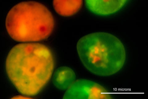
Fluorescent dyes like Fluorescein diacetate can be used to assess viability as the dye passes through the cell membranes, then gets hydrolyzed into fluorescein and appears green in a fluorescent microscope, dead cells appear red. Rhodamine 123 or B can be used to determine mitochondrial membrane potential and respiratory activity. Fluorescent dyes like Acridine orange can be used to study cell division, nuclear dynamics and division. DAPI (4′,6-diamidino-2-phenylindole), is a fluorescent stain that binds strongly to adenine–thymine-rich regions in DNA and Hoechst 33342 are also used to stain the nuclei of both live and fixed cells. See review by A. Sinha (2018) for more information about fluorescent dyes used to investigate cellular processes in yeast cells. Beer can also be studied with a polarizing microscope like the Motic BA310 Pol after beer is frozen or induced to form crystals (Alex 2016, Berdan, 2019).


To view fluorescence requires a fluorescent wide field or confocal fluorescent microscope. Motic offers several fluorescent attachments for their microscopes. E.g. Reflective Epi-Fluorescence attachment [BA400] which integrates into the BA410 arm, the Epi-fluorescence attachment accommodates three filter cassettes for excitation observation
Summary & Conclusions
Yeast cells are essential in the production of beer and wine and a microscope permits the ability to count yeast cells and assess if there might be contaminants for quality control. In research yeast cells are a model system used to study cell division, intracellular trafficking, toxicity and the genomic structure permits the analysis of the cells at the molecular level. Studies are underway to modify the percentage of ethanol yeast can produce for biofuels. The relatively ease at which yeast can be grown and genetically manipulated makes them good models for studying biological properties and test for new drugs. Light microscopy continues to play an important role in the analysis of yeast.
Acknowledgements: I would like to thank Blake Enemark and Jordan Smith for a tour of the Tailgunner brewery in Calgary, some beer samples and information about how they use a microscope to monitor yeast in their facility. Tail Gunner Brewing in Calgary https://www.tailgunnerbrewing.ca/
Related Products
| Models |  |
 |
| Features |
The BA310 is designed for the daily routine work in universities, clinics, laboratories, and life sciences or medical applications. |
Our Moticam A Series cameras are designed with microscopy beginners, teaching environments, hobbyists, and small labs in mind. Learn more> |
Want to know which microscopes fit you best? Send us a message and our specialists are glad to help!
References
YouTube Videos

Industrial and material sciences focus on studying the structure, properties, and performance of materials used in manufacturing, engineering, and product development. These fields involve close...

Microscopes are essential tools in biomedical research. These instruments help scientists, lab professionals, and researchers examine cells, tissues, bacteria, and other microscopic structures that are...

