

Capture Every Detail with Moticam Pro S7 MONO Featuring Sony Pregius Sensor
Motic America
Read Article
Free Ground Shipping on Orders Over $300 Across USA and CANADA
Posted by Motic America on
If you have a freshwater aquarium at home, chances are that you’re familiar with hydras, primitive alienesque critters belonging to the Cnidaria animal group. This group includes other diverse and beautiful creatures such as jellyfish, corals and sea anemones. Cnidarians are among the oldest form of multicellular life known to humanity, with a fossil record reaching back to 700 million years! Most cnidarians occupy marine habitats, but a small fraction can be found in freshwater ponds and lakes, like green and brown hydras. About 11 000 different species form the Cnidaria phylum and all of these species are characterized by specialized cells called cnidocytes. These stinging cells are most abundant on the epitheliomuscular layer of the tentacles and when triggered by a predator or a prey, they explosively unleash their content made of a nematocyst equipped with barbs, spines and a long filament. This nematocyst is what makes Cnidarians effective predators by injecting a paralyzing cocktail of neurotoxins or by entangling their preys.
General biology
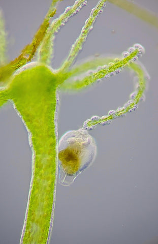
Hydra polyps are amongst the main representatives of freshwater cnidarians and are found in every continent across the globe except for Antarctica. They have been observed living at near freezing temperatures to 25°C and from shallow waters to depts that can reach more than 60 meters. They possess a simple body plan consisting of a head with a combined mouth-anus structure equipped with tentacles, a body column and an adhesive foot, also called pedal disk, that can attach to different substrates such as submerged and floating aquatic plants, dead leaves, sticks, stones and even aquarium windows! Hydras also have the ability to glide freely on substrates aided by their mucous-secreting pedal disk and to float at the surface of the water column by forming

gas bubbles. The body of a hydra is generally around 1 to 20mm in length and is highly contractile; it has the ability to extend up to 30 mm or can rapidly contract in response to light exposure and different chemical or physical stimuli.
Feeding
Freshwater hydras are predominately predators using their stinging cells located on their tentacles to paralyze before consuming a variety of preys such as small crustaceans, annelid worms, insect larvae and small larval fish. However, hydras are being preyed upon themselves by crayfish, flatworms, large amoeba and other animals that can resist being harpooned by their toxic nematocysts.
When waiting for food organisms to pass by, hydras open and extend their tentacles until a prey brush against them. At this point, nematocysts are discharged from the cnidocyte cells and other tentacles may join the attack. The prey is now helpless and will slowly be redirected towards the opening mouth of the hydra. Although, if a prey possesses a hard skeleton and is big enough, it may be able to escape its predator.
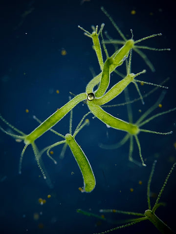
Certain species of Hydrozoans, like Hydra viridissima, commonly named green hydra, have established an endosymbiotic relationship with Chlorella, a green alga that gives the hydra its colour. Chlorella lives within the hydra’s gastrodermal cells, where it produces sugars from sunlight by photosynthesis, and shares some of this precious energy with its host in exchange of shelter and protection.
Reproduction
 Budding is the main asexual reproductive mechanism in freshwater hydras and is very common when food is abundant and environmental conditions are optimal. As seen in the videos, young hydras are able to emerge from the body column of the parent hydra and when mature, they bud off as a free-living polyp. A well-fed hydra is able to reproduce asexually every 3 to 4 days and sometimes multiple buds can appear at the same time on both sides of the body column.
When environmental conditions are harsher, and food is scarce, hydras prefer to reproduce sexually by developing temporary gonads, like testes or ovaries, on the surface of their body column as rounded protuberances instead of buds. In order for eggs in mature female gonads to be fertilized, male sperm must be released into the water. A cyst then forms around the embryo to protect it until environmental conditions are favorable again and it can break free.
The amazing regenerative capacities of hydra’s tissues caught the attention of lots of biologists. If a hydra is cut into small fragments, within two or three days a whole new hydra will emerge from every piece, except those coming from the tentacles or the adhesive foot.
How to View Them?
You can either observe hydras with a compound or stereo microscope. One of the best ways to get contrasting images of brown or green hydras is with a dark background created either by darkfield or oblique illumination. To observe them catching preys and move around, it’s best not to use any cover slip on top of the animal and put a bit of water, just enough to get clear images that aren’t shaky.

The videos and photographs were made using the Motic BA310 compound microscope with oblique illumination and an iPhone 11 Pro mounted on a specialized adaptor.
Related Products
| Models |  |
 |
| Features |
The BA310 is designed for the daily routine work in universities, clinics, laboratories, and life sciences or medical applications. |
Our Moticam A Series cameras are designed with microscopy beginners, teaching environments, hobbyists, and small labs in mind. Learn more> |
Want to know which microscopes fit you best? Send us a message and our specialists are glad to help!
References

Industrial and material sciences focus on studying the structure, properties, and performance of materials used in manufacturing, engineering, and product development. These fields involve close...

Microscopes are essential tools in biomedical research. These instruments help scientists, lab professionals, and researchers examine cells, tissues, bacteria, and other microscopic structures that are...